The Ministry of Health and Child care recently revealed that there are several oral manifestations that are linked to early HIV infections. The oral cavity (mouth) is usually the first place to show signs of HIV infection due to a compromised immune system and is predisposed to various kinds of infections. A�
It is your responsibility, Dear Reader, to take care of your oral health and to know your HIV status.
The following are some of the most common manifestations:
Candidiasis
– Candidiasis is a fungal infection due to any type of Candida. When it affects the mouth, it is commonly called thrush. Signs and symptoms include white patches on the tongue or other areas of the mouth and throat.
– Colonisation and infection of the oral mucosa by Candida species is among the earliest and most common findings in people living with HIV. The oral mucosa is the mucous membrane lining the inside of the mouth.
– Lesions range from white to red or red and white combinations. A lesion is any abnormality in the tissue of an organism, usually caused by disease or trauma.
– The lesions may not be painful or there may be mild discomfort.
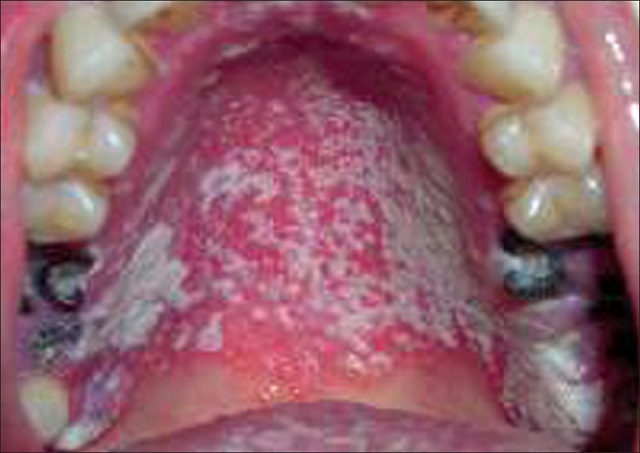
Pseudomembranous Candidiasis (ABOVE)
– White spots or plaques anywhere in the mouth
– Patient may report burning sensation
– Diagnosis usually made upon clinical examination
Erythematous Candidiasis
Erythematous (atrophic) candidiasis is where the condition appears as a red, raw-looking lesion.
– Patchy erythema is usually located on palate and the mid-body (dorsum) of the tongue
– Occasionally on buccal mucosa (The inner lining of the cheeks and lips)
– Patient may report burning sensation
– Usually clinical diagnosis or after response to antifungal therapy
– It may also be a result of toothpaste-induced irritation
Angular Cheilitis
An inflammatory condition that occurs in one or both angles of the mouth.
– Red fissures (cracks) or linear ulcers can be seen.
– These are located at the corners of the mouth.
– It often occurs with intra-oral candidiasis.
Unfortunately, these ulcers can be passed from one person to another through kissing especially when the next person has a weak immune system. Babies are most exposed to such infections if their mothers are also infected.
Kaposia��s sarcoma
Kaposi sarcoma (KS) is a type of cancer in which patches of abnormal tissue grow under the skin or mucous membranes in the mouth
– People living with HIV are vulnerable to a variety of oral malignancies including Kaposia��s sarcoma, malignant lymphoma and squamous carcinoma.
– Kaposia��s sarcoma is the most common.
– The palate is the most common site. In the early stage, the tumor appears as a red to purple bruise.
Hairy leukoplakia
Hairy leukoplakia (also known as oral hairy leukoplakia OHL, or HIV-associated hairy leukoplakia), is a white patch on the side of the tongue with a corrugated or hairy appearance. It is caused by Epstein-Barr virusA� (EBV) and occurs usually in persons who are immuno-compromised (cannot be scraped off.)
– This variety of leukoplakia was first recognised in people who are HIV positive but has been encountered in other immune deficiency states such as organ transplant patients who are intentionally immune suppressed.
– The lateral tongue is the mostA� common location.
– Lesions are of rough texture, adherent and asymptomatic.
– Hairy leukoplakia may be confused with candidiasis. A patient who presents with a white lesion should be treated with antifungal therapy first. If it fails to heal, it most likely is hairy leukoplakia.
Gingival and periodontal lesions
Gingivitis is the mildest form of periodontal disease. It causes the gums to become red, swollen, and bleed easily.
People living with HIV are vulnerable to necrotising gingivitis and periodontitis.
Untreated gingivitis can advance to periodontitis. With time, plaque can spread and grow below the gum line.
Toxins produced by the bacteria in plaque irritate the gums. The toxins stimulate a chronic inflammatory response in which the body in essence turns on itself, and the tissues and bone that support the teeth are broken down and destroyed.
Gums separate from the teeth, forming pockets (spaces between the teeth and gums) that become infected.
Necrotising ulcerative periodontitis is characterised by rapid loss of attachment, connective tissue destruction and deep bone pain.
Linear gingival erythema
– Marginal linear erythema across the attached gingiva generally involving all the teeth
– Gingiva has a bright red appearance
– Patient experiences spontaneous bleeding or bleeding on probing
– Amount of plaque inconsistent with amount of erythema
– No ulceration, no attachment loss seen
– Does not respond to plaque control, scalingA� and root planning
Other conditions associated with HIV in the mouth are Apthous ulcers and Dental caries.
These conditions can be managed with anti-retroviral therapy (ART), oral hygiene instruction and specific treatment of the oral infection.
Get tested for HIV and know your status. Seek treatment early where necessary and take medicines as prescribed by health personnel.